Orfila Rouvière Museum
This museum houses part of the donation made by Paris Descartes University in 2011 from the collections of the former Delmas, Orfila, and Rouvière Anatomical Museums (AMADOR). Among these items are a papier-mâché gorilla by Louis Auzoux and realistic19th-century anatomical wax models, which were displayed in Pierre Spitzner’s traveling museum.

Learn more
A massive donation of over 8,000 items that formed the foundation of the former Delmas-Orfilla-Rouvière Museums (AMADOR)
In 2011, the Faculty of Medicine accepted an exceptional donation: the collections of the formerDelmas-Orfila-Rouvière anatomical museums, formerly located on the premises of the Faculty of Medicine at Paris Descartes University, a collection estimated to contain more than8,000 pieces dating from the19th and20th centuries. With this donation, Montpellier’s anatomical collection now comprises more than13,600 items classified as historic monuments, making it one of the largest in Europe.
Three medical anatomists contributed to the expansion of the Parisian collections during the19th and20th centuries: Mateu Josep Bonaventura Orfila, the physician who founded the anatomy laboratory at the Paris School of Public Health in 1844, followed by Henri Rouvière (1876–1952) and André Delmas (1910–1999), both anatomical pathologists trained at the Faculty of Medicine in Montpellier.
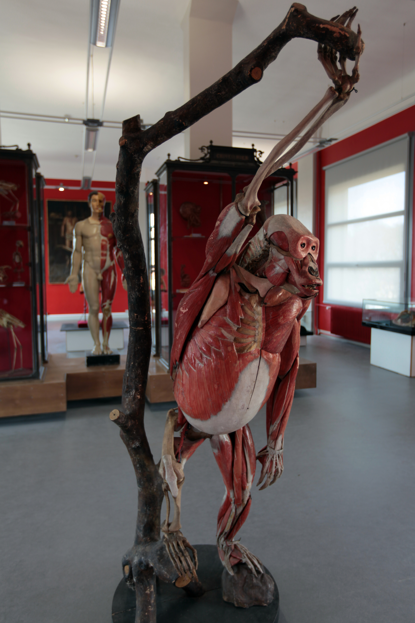
Louis Auzoux's Gorilla
This anatomical model by Dr. Louis Auzoux is a life-size cutaway model that reveals the animal’s full anatomical complexity.
Napoleon III had received a female gorilla as a diplomatic gift from Gabon. After telling the Emperor of his desire to dissect a great ape, Louis Auzoux was fortunate enough to obtain the gorilla—preserved in a barrel of alcohol and transported from Gabon—upon the animal’s death in 1863.
This life-size anatomical model is believed to have been completed between 1866 and 1867.
Glossary:
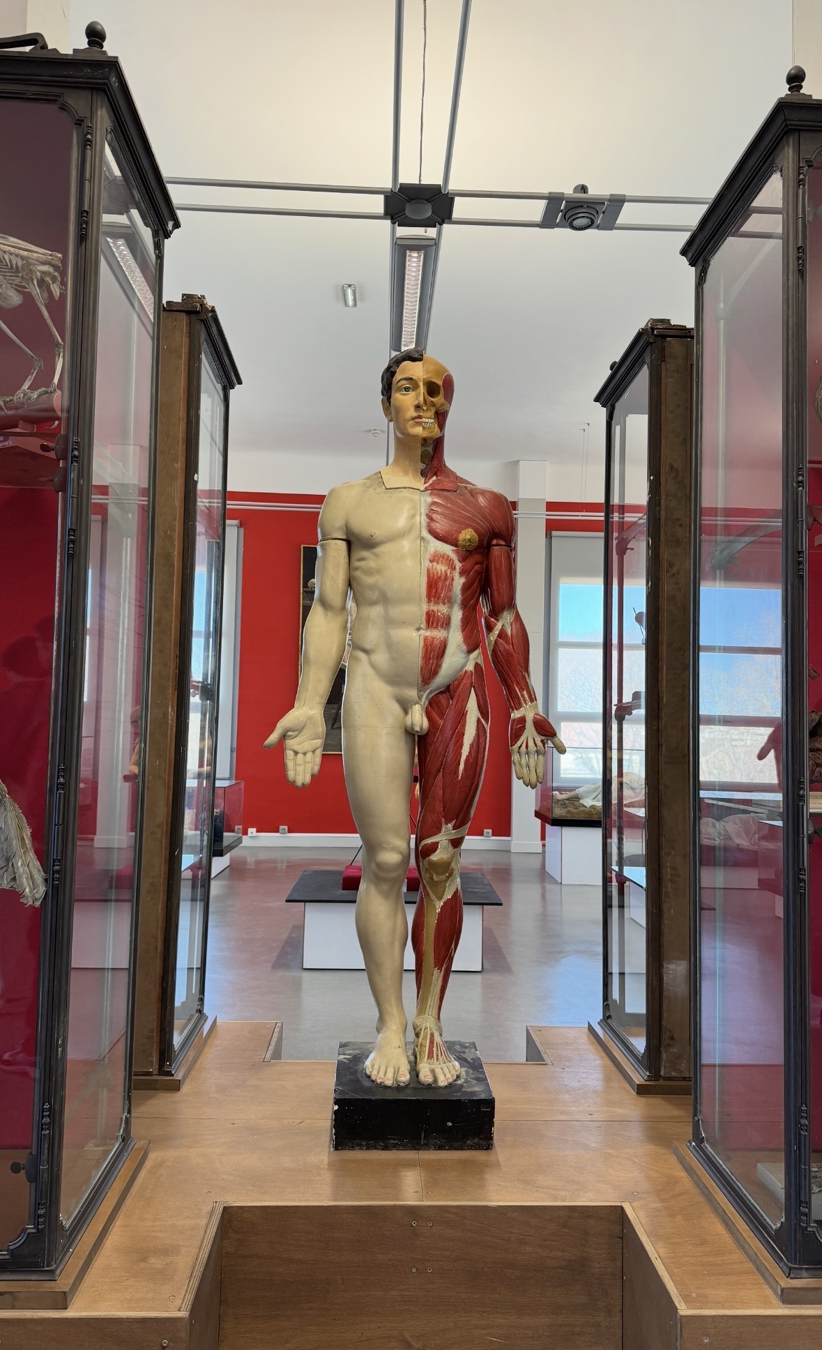
Anatomical model: a model (drawing or sculpture) from which the skin has been removed to reveal the muscles and tendons
Partial flayed model: the skin is removed from only one half of the body, allowing for a comparison between the muscles and tendons on one side and the body’s surface on the other; these partial flayed models were widely used in art schools, helping artists understand the anatomy of the nude figures they were drawing
Clastic model: a disassembled anatomical model that allows users to see how the different anatomical parts fit together
The Spitzner Collection, one of a kind
In addition to this remarkable educational collection is a special collection from thetraveling museum of Pierre Spitzner(1833–1896). In 1856, he founded theGrand Musée Anatomique et Ethnologiquein Paris, which later became atraveling fairground anatomical museum in Northern Europebefore settling permanently in Brussels from the 1920s to the 1960s. Since traveling museums were designed toattract the general public at fairgrounds as profit-driven attractions, some pieces in the collection (299 items) are of a spectacular nature, such as the Venus figures.
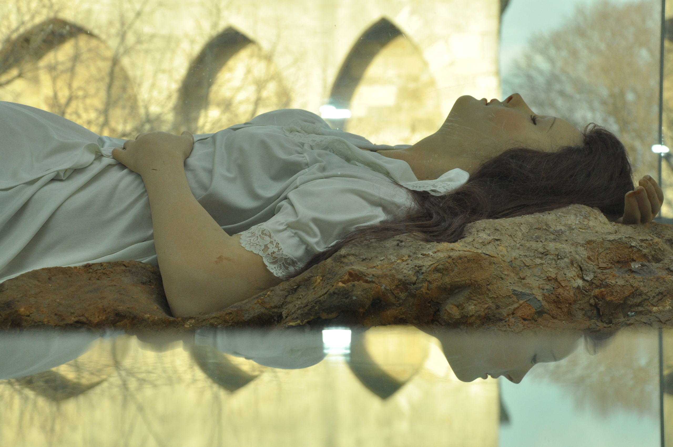
The Sleeping Venus from Pierre Spitzner’s traveling anatomical museum
A wax figure equipped with an artificial respiration mechanism, the Sleeping Venus was used to pique the public’s curiosity and entice them to enter Pierre Spitzer’s fairground booth. 150 years later, she brings to mind the high-fidelity simulation mannequins used today in medical training—realistic mannequins also equipped with a system that mimics breathing.
Spitzner’s fairground booth showcased the latest scientific and medical developments of the time, as well as a broad overview of common diseases titled “Social Hygiene Collection,” for the purposes of moral instruction and education.
Come check out our series on childbirth!
Commentary on the “Anatomical Venuses”: these anatomical models were originally intended for the observation of viscera (internal organs). They are called “Venuses” because the vast majority of them depict female subjects. These Venuses originated in18th-century Italy. Pope Benedict XIV in Bologna, followed by LeopoldI, Grand Duke of Tuscany, in Florence (the future Emperor Leopold II), commissioned these models with the aim of educating the general public about scientific anatomy. This intellectual project was part of the Enlightenment, driven by a scientific goal: to move beyond obscurantism. The Pope in Bologna even asked his clergy to encourage the faithful to donate their bodies to science. It should be noted that these models were never intended for the education of doctors, as they had access to actual dissection sessions. Historians hypothesize that these Venuses were most often women because feminine beauty held particular importance in Italian art. Moreover, it was important that beauty characterized these models to soften the impact of observing the perfectly realistic—and rather unsavory—internal organs.